
Ultrastructural alterations and mitochondrial dysfunction in skeletal muscle of peripheral artery disease patients
implications for early therapeutic interventions
DOI:
https://doi.org/10.17179/excli2024-7592Keywords:
peripheral artery disease, mitochondria, sarcomere atrophy, intramyocellular lipids, sarcoplasm, muscleAbstract
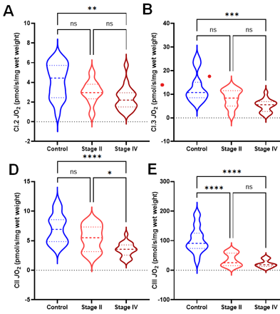
Peripheral artery disease (PAD) is an atherosclerotic condition that impairs blood flow to the lower extremities, resulting in myopathy in affected skeletal muscles. Improving our understanding of PAD and developing novel treatment strategies necessitates a comprehensive examination of cellular structural alterations that occur in the muscles with disease progression. Here we aimed to employ electron microscopy to quantify skeletal muscle ultrastructural alterations responsible for the myopathy of PAD. Fifty-two participants (22 controls, 10 PAD Stage II, and 20 PAD Stage IV) were enrolled. Gastrocnemius biopsies were obtained to determine mitochondrial respiration and oxidative stress. Skeletal muscle sarcomere, mitochondria, lipid droplets, and sarcoplasm were assessed using transmission electron microscopy and focused ion beam scanning electron microscopy. Controls and PAD Stage II patients underwent walking performance tests: 6-minute walking test, 4-minute walking velocity, and maximum graded treadmill test. We identified several prominent ultrastructural modifications in PAD gastrocnemius, including reduced sarcomere dimensions, alterations in mitochondria number and localization, myofibrillar disorientation, changes in lipid droplets, and modifications in mitochondria-lipid droplet contact area. These changes correlated with impaired mitochondrial respiration and increased ROS production. We observed progressive deterioration in mitochondrial parameters across PAD stages. Stage II PAD showed impaired mitochondrial function and structure, while stage IV exhibited further deterioration, more pronounced structural alterations, and a decrease in mitochondrial content. The walking performance of Stage II PAD patients was significantly reduced. Our findings suggest that pathological mitochondria play a key role in the skeletal muscle dysfunction of PAD patients and represent an important target for therapeutic interventions aimed at improving clinical and functional outcomes in this patient population. Our data indicate that treatments should be implemented early and may include therapies designed to preserve and enhance mitochondrial biogenesis and respiration, optimize mitochondrial-lipid droplet interactions, or mitigate oxidative stress.
Downloads
Additional Files
Published
How to Cite
License
Copyright (c) 2024 Dylan Wilburn, Emma Fletcher, Evlampia Papoutsi, William T. Bohannon, Gleb Haynatzki, Bernd Zechmann, Yuqian Tian, Iraklis Pipinos, Dimitrios Miserlis, Panagiotis Koutakis

This work is licensed under a Creative Commons Attribution 4.0 International License.
Authors who publish in this journal agree to the following terms:
- The authors keep the copyright and grant the journal the right of first publication under the terms of the Creative Commons Attribution license, CC BY 4.0. This licencse permits unrestricted use, distribution and reproduction in any medium, provided that the original work is properly cited.
- The use of general descriptive names, trade names, trademarks, and so forth in this publication, even if not specifically identified, does not imply that these names are not protected by the relevant laws and regulations.
- Because the advice and information in this journal are believed to be true and accurate at the time of publication, neither the authors, the editors, nor the publisher accept any legal responsibility for any errors or omissions presented in the publication. The publisher makes no guarantee, express or implied, with respect to the material contained herein.
- The authors can enter into additional contracts for the non-exclusive distribution of the journal's published version by citing the initial publication in this journal (e.g. publishing in an institutional repository or in a book).


